StudentShare
Contact Us
Sign In / Sign Up for FREE
Search
Go to advanced search...
Free

The Basic Principles of Magnetic Image Resonance Production - Essay Example

Cite this document
Summary
The researcher of this essay aims to analyze magnetic resonance imaging (MRI), that is a radio diagnostic tool that has revolutionized medical imaging. This is a modest attempt to bring out the basic physics, instrumentation and imaging principles used in MRI…
Download full paper File format: .doc, available for editing
GRAB THE BEST PAPER91.3% of users find it useful
The Basic Principles of Magnetic Image Resonance Production
Read Text Preview

Extract of sample "The Basic Principles of Magnetic Image Resonance Production"

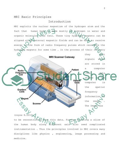
 MRI Basic Principles Abstract Magnetic resonance imaging (MRI) is a radio diagnostic tool that has revolutionized medical imaging. This is a modest attempt to bring out the basic physics, instrumentation and imaging principles used in MRI MRI Basic Principles Introduction MRI exploits the nuclear magnetism of the hydrogen atom and the fact that human body is made mostly of hydrogen in water and organic molecules like fats. These tiny hydrogen magnets can be aligned along external magnetic fields and can be made to receive energy in the form of radio frequency pulses which reorients the hydrogen magnets for some time . In the process of their recovery they emit signals which are stored in a computer .The data stored in the computer is the spatial frequency information of the image . The Image has (coyne K.2010) to be reconstructed from this data. Further locating a slice of the human body along different axis also need complicated instrumentation . Thus the principles involved in MRI covers many disciplines like physics , engineering, image processing and medicine. MRI How it works The Components of MRI(Mcrobbie etal ,2006)are The magnet Which produces a static field of the order of one Tesla. Patient couch is through the bore of this magnet .The magnetic field is along the axis of the bore. The hydrogen nuclei in the tissues of the patient try to align along the magnetic field and in the process end up with their spins rotating about the direction of the magnetic field .This motion is called Larmor precession and has a frequency γBo Mhz where γ is called the gyromagnetic ratio which has a value 42.57 for hydrogen and Bo is the magnetic field .Hence for a one Tesla field the Larmor frequency is 42.57Mhz RF coil RF coils are used to transmit MR signals of the Larmor frequency to the tissues and receive from the tissues MR signals containing diagnostic information .The RF coils surround either whole or part of the body. When RF pulses are applied for a short period the rotation axis of the spin nuclei changes and sometime after the end of the pulse the spins reorient .During this reorientation the receiver coils picks up signals from the tissues which carries information about the tissues Gradient coils Gradient coils are used for providing pulses which can add to or subtract from the static field there by providing a magnetic field gradient along three perpendicular directions. The linearly changing magnetic field gives rise to a proportional larmor frequency. This helps in locating slices along the three directions by tuning the frequency. The Gradient coils are placed inside the bore of the magnet Computers The Gradient pulses select the slices and the tuned RF pulses are transmitted periodically .The MR signal received from the tissues contain spatial frequency information which are called K-space data .This is stored in computers and using softwares the image is reconstructed by finding the inverse Fourier transform of the K-space data Image Production The first step in image production is selecting the slice .This is achieved by sending a appropriate Gradient pulse at the same time as the RF pulse .The Gradient pulse determines the Larmor frequency of the selected slice and hence the frequency bandwidth of the RF signal. The selected slice is like the field of view of a camera (FOV) and the MR signal received is scanned in terms of frequency(Frequency Encoding) along one direction and in terms of the phase(phase encoding) along the other .The number of samples determines the size of the computer memory required to store the image. Each location corresponds to a particular frequency and a particular phase .The data stored is the location having that particular frequency an phase. Such data is called K-space data and has to be transformed to real picture by complex computer programs(WestBrook and Carolyn 2000 ,McRobbie etal 2006) Basic MRI scans The Timing parameters The effect of the static magnetic field B is to make the magnetic moment of the hydrogen nuclei to precess about the direction of B which we take as the longitudinal axis .By applying an RF pulse of larmor frequency the axis of precession can be made to rotate through 90 degrees. The precession now is about a transverse axis. As the magnetic moment rotates about ` Source:(McRobbie etal 2006) the transverse axis a voltage is induced in the receiver coil. As the RF pulse is switched off the transverse magnetization decreases and this is called decay . At the same time there is an increase in longitudinal magnetization which is called recovery. TR is the minimum time interval between two RF pulses TE is the time interval between the application of RF pulses and receiving the MR signal in the receiver coil T1 is the recovery time after the application of RF T2 is the decay time after the application of the RF(2) T1 and T2 depend upon the tissue types. Fluids have long T1.Water has medium and fat has very short T1 .T2 tends to be shorter than T1. Fluids has long, water medium and fat minimum T2(McRobbie etal 2006). Image contrast Different types of tissues has different intensities in the MRI image which is called the image contrast. The image contrast depend on the proton density PD and the timings T1 and T2.Depending on the signal times we have three different types of images Short TR and TE gives T1 weighted images. In T1 weighted images fluids are very dark ,water is mild grey and fat is intense. Long TR and TE gives T2 weighted images .For T2 weighted images fluids has maximum intensity and water and are mild grey(McRobbie etal 2006) Manipulating timing parameters for better contrast All MR images are produced using pulse sequences stored in the scanner computer .The sequence contains RF pulses and gradient pulses .The duration and timing of these pulses determine the image contrast. There are two types of pulse sequences .Spin Echo (SE) and Gradient Echo (GE) sequences. SE uses two RF pulses to produce the echo where as GE uses only one.Both of them can produce T1,T2 and PD weighted images. GE echo also depends on the in-homogeneity of the magnetic field .The combined effect of T2 and in-homogeneity is called T2*.(McRobbie etal 2006) The following table summarizes the effects of timing parameters on contrast. Timing Contrast in sSequences TR TE fluids water fat T1 Weighted SE or GE short short Dark Mid-gray intense T2 weighted SE or GE Long Long intense Mid-gray Dark PD weighted GE Long short Mild gray Mild gray Mild gray MRI T1 and T2 weighted images T1,T2 and PD weighted images (http://www.grin.com/en/doc/281950/linear-spectral-unmixing-approaches-to-magnetic-resonance-image-classification) Image Acquisition The first step in image acquisition is selecting a slice. The slice selectin is achieved by the gradient pulses . An z gradient pulse selects an axial slice, y Gradient an coronal slice and x gradient a sagittal slice. (source :Westbrook and kaut 2000) Once a slice is selected the signal coming from it is received using frequency and phase encoding. The frequency gradient is switched when the signal is received and is called the readout signal. Phase gradient is switched on to localize along the remaining axis. The data thus obtained is in K-space with frequency as one dimension and phase as the other .K-space data is stored in computer memory as a matrix the size of which is determined by the sampling rate. The real image is obtained from k-space data by calculating The fourier transform Conclusion MRI is the best and safest way to see inside the human body. MRI is able to differentiate between the different soft tissues in the body . It also produces images of sections along different axis.It also does not make use of or produce any harmful radiations. A good understanding of MRI principles requires knowledge in Physics ,Engineering ,Imaging and Medicine References Bushong S.(1996) Magnetic Resonance Imaging.Physical and Biological principles Mosbey:ST.Louis:MO Cordoza J & Herfkens R.(1994) MRI Survival Guide : Lippincott Raven :Newyork ,NY Coyne K.2010, MRI:A Guided Tour,National High magnetic Field Laboratory http://www.magnet.fsu.edu/education/tutorials/magnet academy/mri/fullarticle.html English P.& Moore C.(1995)MRI for Radiographers Springer Verlag :London Hashemi R.H. & Bradley W.G.Jr.(1997)MRI The Basics Willaims and Wilkins Baltimore MD Kaut C.(1992) MRI Workbook for Technologists Lippincott Raven :Newyork NY Kaut C.(1994) Review questions for MRI Blackwell Science Oxford Mattson J. and Simon M (1996)The Poineers of NMR and Magnetic Resonance in Medicine.The Story of MRI McRobbie W.Donald, Moore A.Elizabeth,Graves J.Martin and Prince Martin R. 2006 MRI from picture to proton Cambridge University press Jericho NY:Dean Books Co Ness Aiver(1996) All you really need to know about MRI Physics University of Maryland:Baltimore Werhli F.(1991) Fast scan magnetic resonance Principles And applications Raven Press Newyork NY Westbrook C.(1994)Handbook of MRI Technique Blackwell Science ,Oxford Westbrook Catherine, Kaut Carolyn 2000 MRI in practice Blackwell science London Wheeler G. & Withers K.(1996)Magnetic Resonance Imaging and Review Lippincott Raven NY Read More
Cite this document
  • APA
  • MLA
  • CHICAGO
(MRI Basic Principles Assignment Example | Topics and Well Written Essays - 1250 words, n.d.)
MRI Basic Principles Assignment Example | Topics and Well Written Essays - 1250 words. https://studentshare.org/medical-science/1757079-the-basic-principles-of-magnetic-image-resonance-production
(MRI Basic Principles Assignment Example | Topics and Well Written Essays - 1250 Words)
MRI Basic Principles Assignment Example | Topics and Well Written Essays - 1250 Words. https://studentshare.org/medical-science/1757079-the-basic-principles-of-magnetic-image-resonance-production.
“MRI Basic Principles Assignment Example | Topics and Well Written Essays - 1250 Words”. https://studentshare.org/medical-science/1757079-the-basic-principles-of-magnetic-image-resonance-production.
  • Cited: 0 times
sponsored ads
We use cookies to create the best experience for you. Keep on browsing if you are OK with that, or find out how to manage cookies.
Contact Us